

Abstract
Backgrounds:Primary immune thrombocytopenia (ITP) is an acquired autoimmune disease characterized by reduced platelet count and an increased risk of bleeding. The imbalance of Treg/Th17 cells has been demonstrated in ITP, but the mechanism of Th17/Treg cells imbalance is still not clear. In this study, we aimed to investigate whether the expression of helper T (Th) or Treg cell-related microRNAs, such as miR-183-96-182 cluster, miR-17-5p, miR-99a, miR-146-5p, miR-155-5p, miR-181-5p, and miR-326, regulates the ratio of Th17/Treg in CD4+ T cells and could be used to evaluate the clinical implications of ITP patients.
Methods: Peripheral blood was obtained from 54 patients with active ITP and 34 healthy controls. Peripheral blood mononuclear cells (PBMCs) were isolated using Ficoll density-gradient centrifugation and the CD4+ cells were separated by immuno-magnetic microbeads selection. Amplification technique of RT-PCR using stem-loop primers was applied to detect the relative expression of microRNAs (miR-17-5p, miR-99a, miR-96-5p, miR-146a-5p, miR-155-5p, miR-181a-5p, miR-182-5p, miR-183-5, miR-326) and U6 was normalized as control for miRNA quantification. The frequencies of Th17 and Treg cells in peripheral blood were analyzed by flow cytometry. The mRNA expression levels of Il-6, Il-10, Il-17, Rorγ-t and Foxp-3 in CD4+ cells were determined by RT-PCR. Platelet autoantibodies specific for GPIIb/IIIaor GPIb/IX were measured using MAIPA method. CD4+ cells were transfected with miRNAs (miR-99a, miR-182-5p, miR-183-5), mimics or inhibitors, which were used to detect the function of miRNAs. Cytokines in culture medium were determined by ELISA.
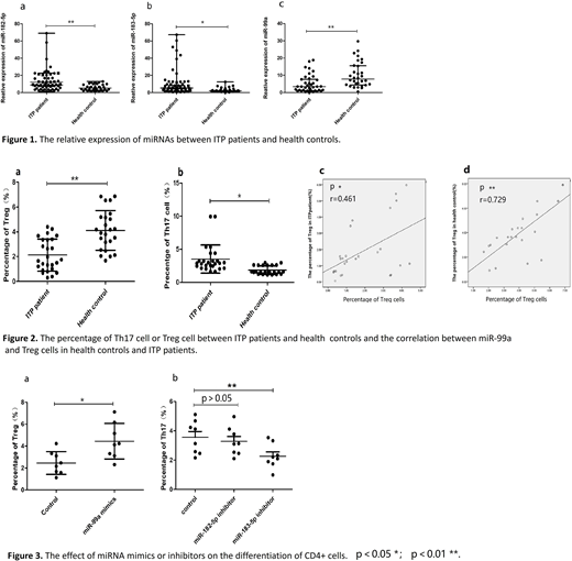
Results: Our results showed that the relative expression of miR-182-5p and miR-183-5p in CD4+ cells was significantly increased in active ITP patients, compared to healthy controls (miR-182-5p, median 9.2678 vs 5.2723, p < 0.05, Fig. 1a; miR-183-5p, median 5.4435 vs 2.009, p < 0.05, Fig. 1b). In addition, the relative expression of miR-99a in ITP patients was lower than that of healthy controls (median 3.4214 vs 7.9648, p < 0.05; Fig. 1c). Moreover, the frequency of Treg cells decreased significantly in ITP patients compared to those in controls (1.89±1.59% vs 4.12±1.42%, p < 0.05; Fig. 2a), and the percentage of Treg cells was positively correlated with the relative expression of miR-99a in ITP patients(r=0.461, p< 0.05; Fig. 2c) and health controls(r=0.729, p< 0.05; Fig. 2d). Though the percentage of Th17 cells increased in ITP patients compared to the health controls (3.51±2.13%vs 1.85±0.63%, p < 0.05; Fig. 2b), there was no correlation between the percentage of Th17 and the relative expression of microRNAs in ITP patients or health controls. Besides, there was no correlation between the expression of mRNAs (Il-10, Il-17, Rorγ-t and Foxp-3) and microRNAs (miR-99a, miRNA-182-5p or miR-183-5p). No significant correlation was found between the microRNAs expression and platelets counts or different autoantibody subsets in ITP patients. The relative expression of other microRNAs (miR-17-5p, miR-96-5p, miR-146a-5p, miR-155-5p, miR-181-5p, miR-326) revealed no difference in CD4+ cells between ITP patients and health controls. Furthermore, the down-regulated expression of miR-183-5p with inhibitors promoted to the differentiation of Th17 cells(Fig. 3a), while up-regulated expression of miR-99a with mimics contributed to Treg cells in CD4+ cells from ITP patients (Fig. 3b). Meanwhile, the IL-17A in culture medium decreased in inhibitor group of miR-183-5p or miR-183-5p. However, miR-182-5p inhibitor had no effect on the differentiation of Th17 cells.
Conclusions: Our results show the abnormal expression of microRNAs (miR-99a, miRNA-182-5p and miR-183-5p) in CD4+ cells and the miR-99a was closely correlated with the Treg cells. The aberrant expression of microRNAs may contribute to the imbalance of Th17/Treg cells in the development of ITP patients and potentially constitute a novel therapeutic target.
No relevant conflicts of interest to declare.

This feature is available to Subscribers Only
Sign In or Create an Account Close Modal